Normal and ineffective erythropoiesis

Erythropoiesis is a vital, life-long process where haematopoietic stem cells differentiate and mature into red blood cells.1 Although a continuous process, erythropoiesis can be thought of as occurring in two stages: early versus late stage.

Normal and ineffective erythropoiesis

Erythropoiesis is a vital, life-long process where haematopoietic stem cells differentiate and mature into red blood cells.1 Although a continuous process, erythropoiesis can be thought of as occurring in two stages: early versus late stage.

Chronic anaemia and ineffective erythropoiesis

Anaemia, a condition that affects almost 25% of the global population, refers to the shortage of functional haemoglobin or reduction in red blood cells.1,2 Anaemia reduces the delivery of oxygen to tissues, which is compensated primarily by increasing cardiac output; the resulting tissue hypoxia can affect the function of major organs.3 Chronic, severe anaemia is associated with increased risk of death due to cardiac failure.4,5 Reduced delivery of oxygen to tissues also causes other symptoms such as fatigue, dyspnoea, tachycardia, hypotension, low body temperature and enlarged spleen.6

The main cause of anaemia is iron deficiency, closely followed by anaemia associated with chronic disease.6 This website will discuss and focus on anaemia associated with haematological diseases.

In anaemia associated with chronic haematological diseases, reduced red blood cell production may occur due to ineffective erythropoiesis. Ineffective erythropoiesis causes an imbalance in the various progenitor and precursor cells that proliferate, differentiate and mature into red blood cells, leading to impaired erythropoiesis.7-11 Ineffective erythropoiesis is a hallmark of certain haematological diseases such as:7-10,12-16

  • myelodysplastic syndromes
  • thalassaemias (e.g. specific types of α- and β-thalassaemia)
  • aplastic anaemia
  • myelofibrosis
  • sickle cell anaemia
  • congenital dyserythropoietic anaemias
  • inherited sideroblastic anaemias.

Please see Chronic anaemia in haematological diseases for further details

What is erythropoiesis?

Erythropoiesis is a vital, complex, life-long process where haematopoietic stem cells proliferate, differentiate and mature into red blood cells.17 Erythropoiesis is a continuous process that can be conceptually divided into early and late stages.18

Early- versus late-stage erythropoiesis

Early-stage erythropoiesis is characterised by proliferation of early-stage erythroid cells (progenitors), whereas late-stage erythropoiesis involves the differentiation and maturation of late-stage erythroid cells (precursors).18

late-stage
late-stage

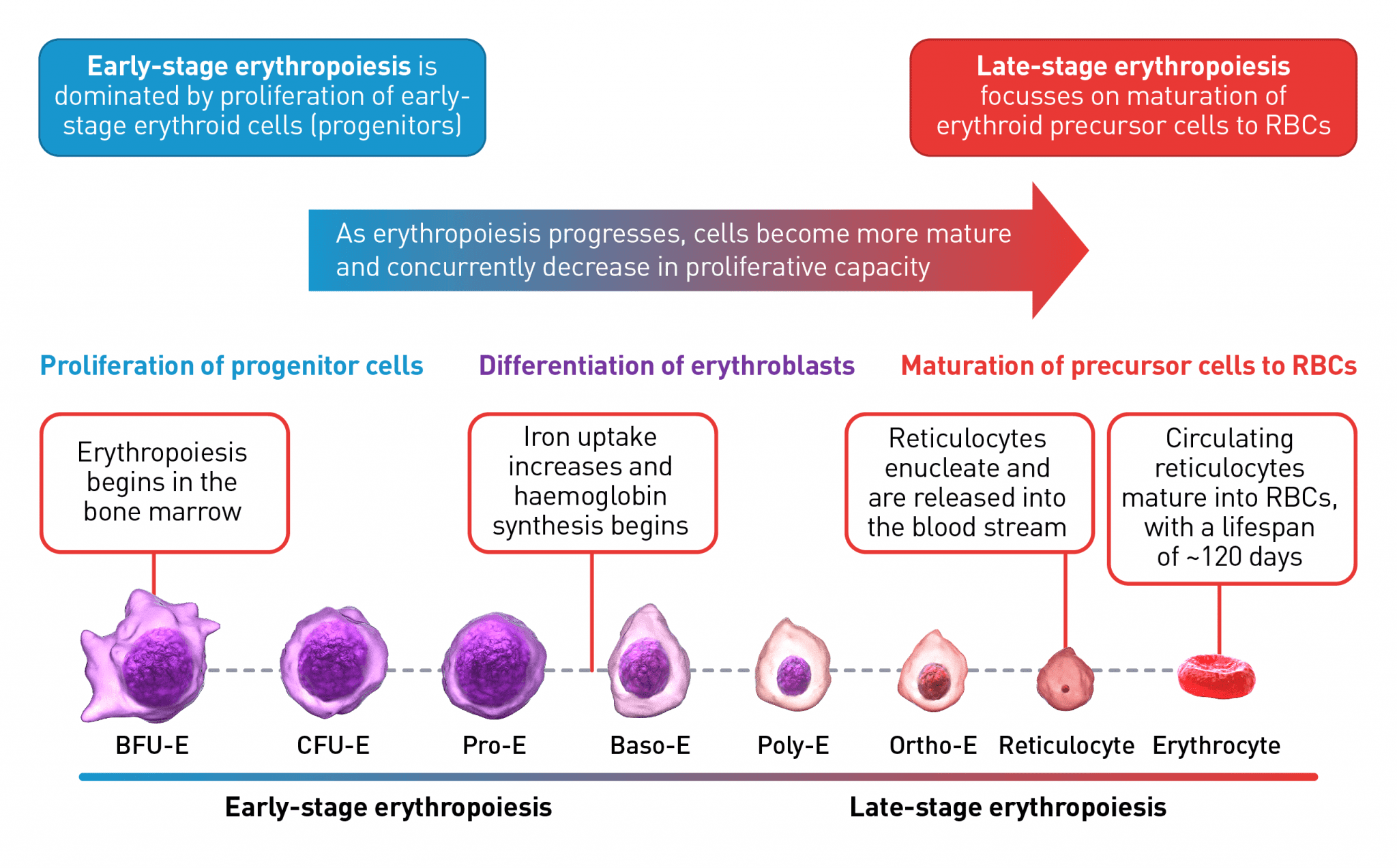
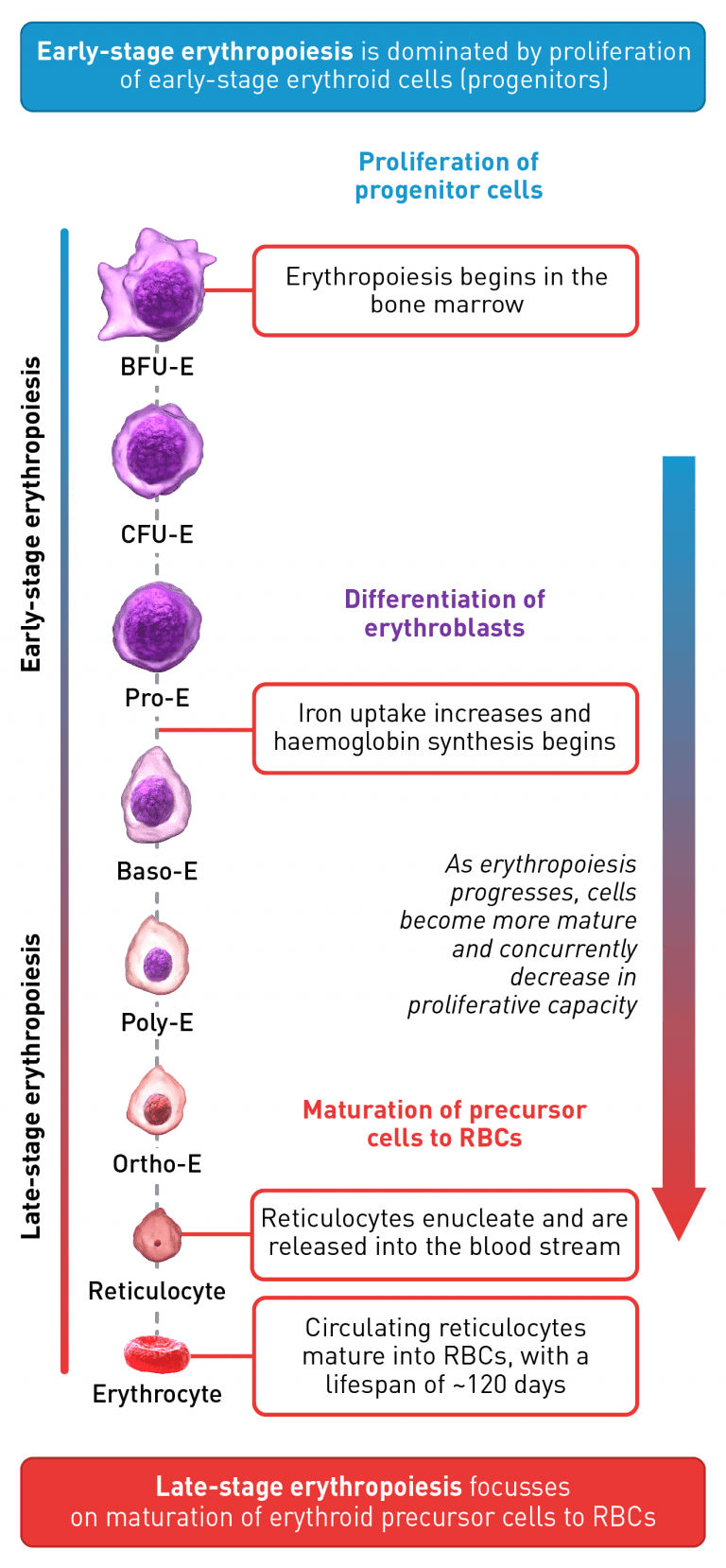
The erythroid progenitor cell compartment contains the early erythroid progenitors. BFU-E represent the earliest progenitors committed exclusively to erythroid maturation, which differentiate into late CFU-E and proerythroblasts.19 The earliest recognisable erythroid cells are the Pro-E, which undergo morphological changes such as reduction in cell size, protein production including haemoglobin, and reduction in proliferative capacity, evolving through the erythroblast stages, Baso-E, Poly-E and Ortho-E, successively.20,21 At the end of the terminal maturation, erythroblasts expel their nuclei and lose all their organelles resulting in mature enucleated cells, called reticulocytes.20 After expelling its nucleus, the reticulocyte is released into the bloodstream where maturation continues in order to produce fully functional, biconcave erythrocytes within 1–2 days.20

Baso-E, basophilic erythroid; BFU-E, burst-forming unit-erythroid; CFU-E, colony-forming unit-erythroid; Ortho-E, orthochromatophilic; Poly-E, polychromatophilic; Pro-E, proerythroblasts; RBC, red blood cell.

The process is tightly regulated by many different transcription factors and signalling pathways, as shown in the figure below.13,18,22-30

fig 2
fig 2

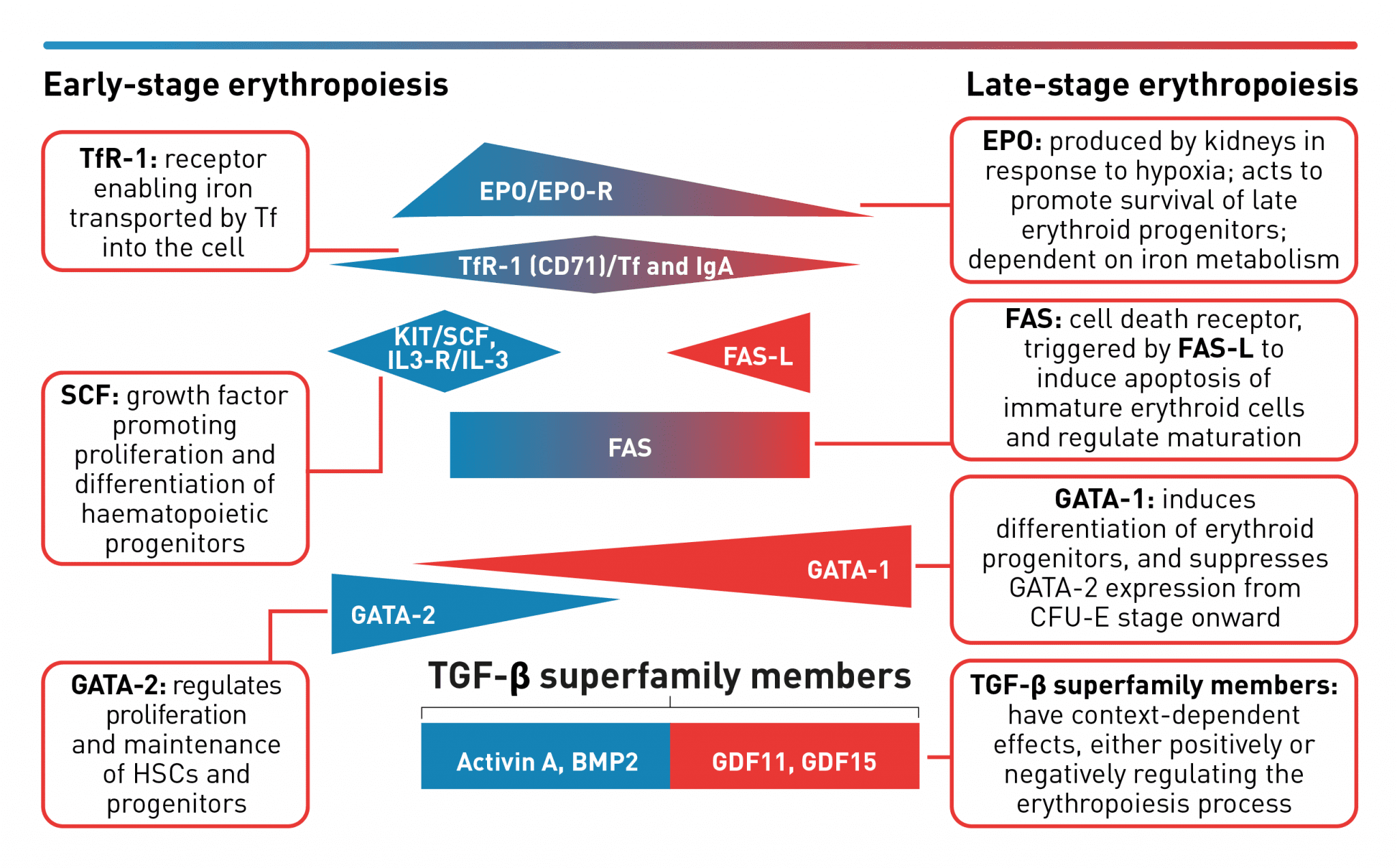
A complex network of transcription factors and epigenetic regulators ensure appropriate numbers of red blood cells are produced.18,31,32 EPO promotes survival and proliferation of erythroid progenitor cells by preventing apoptosis.33 GATA-1 is the main regulator of lineage-commitment, differentiation and survival of early-stage erythroid cells18,31 Other transcription factors include regulators of iron metabolism, such as TfR1 and early acting haematopoietic growth factors such as SCF.18 FAS-L is expressed by late-stage erythroid cells to trigger apoptosis of immature erythroid cells and regulate maturation, while selected TGF-β superfamily ligands also play a key role in regulating erythroid maturation.18,30,32,34,35 Tight regulation of erythropoiesis is crucial to ensure a steady-state process.18

BMP2, bone morphogenetic protein 2; EPO, erythropoietin; CFU-E, colony-forming unit-erythroid; EPO-R, erythropoietin receptor; FAS-L, FAS ligand; GDF, growth differentiation factor; HSC, haematopoietic stem cells; IL-3, interleukin 3; IL-3-R, interleukin 3 receptor; IgA, immunoglobulin A; SCF, stem cell factor; Tf, transferrin; TfR-1 (or CD71), transferrin receptor; TGF, transforming growth factor.

Erythropoietin stimulates red blood cell production by acting as a key positive regulator of early-stage erythropoiesis; its greatest influence occurs during the colony-forming unit-erythroid (CFU-E)/proerythroblasts stage of erythropoiesis and wanes during the later maturation stages.13,33,36,37 The transcription factor GATA-2 is highly expressed in haematopoietic stem cells and early-stage progenitors, but its expression is suppressed from the CFU-E stage and beyond.38 Expression of GATA-1 is first seen in CFU-E progenitors and increases as the erythroblasts differentiate and mature. This transition of GATA expression from GATA-2 to GATA-1 during the erythropoiesis process, as indicated in the figure above, is referred to as the GATA switch mechanism, and results in the mutually exclusive expression of GATA-2 and GATA-1 during early and late stages of erythropoiesis, respectively.23,38, FAS ligand is expressed by late-stage erythroid precursor cells and is involved in regulating the final maturation stages that lead to the generation of functional red blood cells.18 Members of the transforming growth factor β (TGF-β) superfamily can exert opposing regulatory effects on the erythropoiesis process.30 Signalling mediated by selected TGF-β superfamily ligands regulates erythroid maturation via the Smad pathway; dysregulation of this signalling pathway may contribute to impaired erythroid maturation leading to ineffective erythropoiesis.34,39,40

Normal versus ineffective erythropoiesis

In healthy individuals, normal erythropoiesis maintains the circulating erythrocyte count within remarkably narrow limits.11,13,33 Successful erythropoiesis is dependent on maintaining a tightly regulated differentiation pathway to retain appropriate levels of erythrocytes. In ineffective erythropoiesis, there is an imbalance in erythropoiesis through increased proliferation of erythroid progenitors accompanied by increased apoptosis a nd reduced maturation of erythroid precursors.7-11 Ineffective erythropoiesis is a prominent component of the anaemia that accompanies a range of haematological diseases, including myelodysplastic syndromes, β-thalassaemia, and congenital dyserythropoietic anaemias.12

Characteristics of ineffective erythropoiesis

Haematological disorders are often associated with alterations in certain signalling pathways that regulate erythropoiesis.8,9,11,34,39,41-43

Please see Erythroid maturation defect (EMD) for further details

fig 3
fig 3

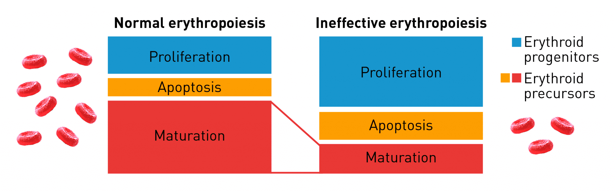
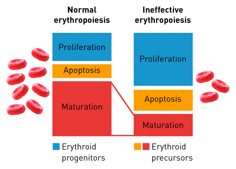
Normal erythropoiesis proceeds with maturation and apoptosis tightly regulated to maintain red blood cell homeostasis and normal oxygen levels. Ineffective erythropoiesis is an ongoing pathological state in which increased erythroid proliferation cannot restore red blood cell count due to increased apoptosis of immature erythroid cells and impaired late-stage maturation.11

Implications of ineffective erythropoiesis

Additional to the range of symptoms associated with the chronic anaemia seen in haematological diseases, such as cardiac failure, fatigue, dyspnoea, tachycardia, hypotension, low body temperature and enlarged spleen, ineffective erythropoiesis may also cause iron overload, which has its own complications.4,6,43,44 Iron, bound to plasma transferrin, circulates the body and accumulates within cells in the iron-storage protein ferritin. Hepcidin regulates iron metabolism by inhibiting iron absorption in the gut. Ineffective erythropoiesis suppresses hepcidin production in the liver, leading to unrestrained intestinal iron uptake.18,43,45 Additionally, the body attempts to compensate for the haemoglobin shortage by absorbing more iron. Increased iron levels result in saturation of transferrin and, as there is no physiological pathway to excrete iron, non-transferrin-bound iron begins to accumulate in the body. This non-transferrin-bound free iron is toxic and as levels in the blood rise, iron deposits in various organs leading to a range of clinical complications including pituitary damage, hyperthyroidism, liver disease, heart failure and diabetes.44,47-53

Please see Treating anaemia in haematological diseases for further details

Ineffective erythropoiesis can also lead to erythroid marrow expansion, which can result in bone deformities in the skull and face, a complication observed in patients with thalassaemias.54,55 Hypercoagulable state, which is linked to a high prevalence of thromboembolic and cerebrovascular events, is also associated with ineffective erythropoiesis.54

fig 4
fig 4

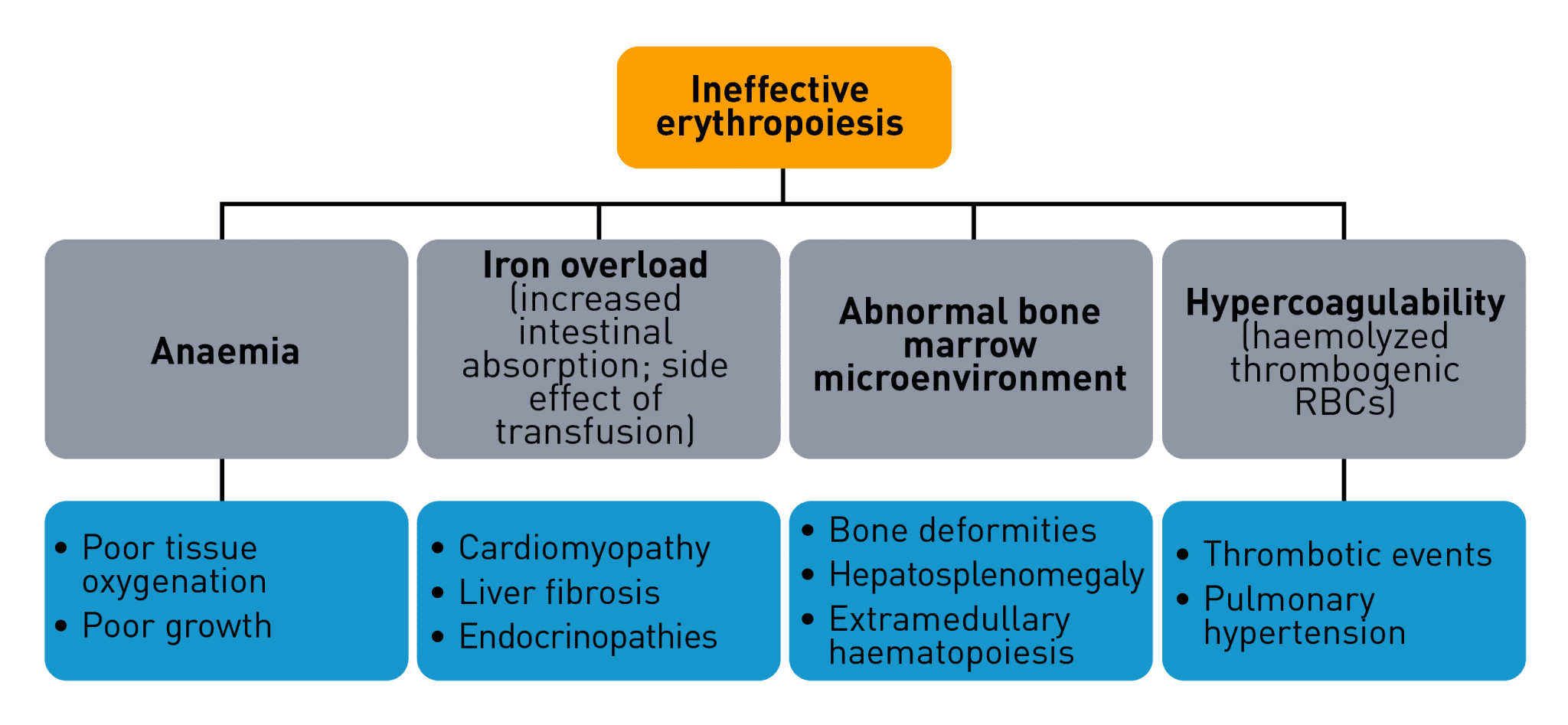
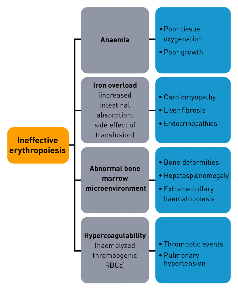
Ineffective erythropoiesis contributes to a variety of symptoms and complications that are characteristic of certain haematological disorders.54-56

RBC, red blood cell.

References

  1. World Health Organization, Edited by, de Benoist B, McLean E, Egli I, Cogswell M. Worldwide prevalence of anaemia 1993-2005: Who global database on anaemia. 2008. Available at: whqlibdoc.who.int/publications/2008/9789241596657_eng.pdf.
  2. Kassebaum NJ, Collaborators GBDA. The global burden of anemia. Hematol Oncol Clin North Am 2016;30:247-308.
  3. Tsai AG, Hofmann A, Cabrales P, Intaglietta M. Perfusion vs. oxygen delivery in transfusion with “fresh” and “old” red blood cells: The experimental evidence. Transfus Apher Sci 2010;43:69-78.
  4. Della Porta MG, Malcovati L, Strupp C, et al. Risk stratification based on both disease status and extra-hematologic comorbidities in patients with myelodysplastic syndrome. Haematologica 2011;96:441-9.
  5. Mozos I. Mechanisms linking red blood cell disorders and cardiovascular diseases. Biomed Res Int 2015;2015:682054.
  6. Lambert J-F, Beris P. Pathophysiology and differential diagnosis of anaemia. In: Beaumont C, Beris P, Beuzard Y, Brugnara C, eds. The handbook disorders of erythropoiesis, erythrocytes, and iron metabolism. Chapter 4. 2009:108-41.
  7. Taher AT, Weatherall DJ, Cappellini MD. Thalassaemia. Lancet 2018;391:155-67.
  8. Bowen D. What is ineffective erythropoiesis in myelodysplastic syndromes? Leuk Lymphoma 1995;18:243-7.
  9. Raj K, Mufti GJ, Hoffbrand VA, Higgs DR, Keeling DM, Mehta AB. The myelodysplastic syndromes. Postgraduate haematology, seventh edition: John Wiley & Sons Ltd.; 2016:438-73.
  10. Gupta R, Musallam KM, Taher AT, Rivella S. Ineffective erythropoiesis: Anemia and iron overload. Hematol Oncol Clin North Am 2018;32:213-21.
  11. Oikonomidou PR, Rivella S. What can we learn from ineffective erythropoiesis in thalassemia? Blood Rev 2018;32:130-43.
  12. Camaschella C, Nai A. Ineffective erythropoiesis and regulation of iron status in iron loading anaemias. BJH 2016;172:512-23.
  13. Higgs DR, Noemi R, Hay D, et al. Erythropoiesis. Postgraduate haematology, seventh edition: John Wiley & Sons Ltd.; 2016:11-20.
  14. Young NS. Aplastic anemia. N Engl J Med 2018;379:1643-56.
  15. Dong X, Han Y, Abeysekera IR, Shao Z, Wang H. GDFf11 is increased in patients with aplastic anemia. Hematology 2019;24:331-6.
  16. Tefferi A. Primary myelofibrosis: 2019 update on diagnosis, risk-stratification and management. Am J Hematol 2018;93:1551-60.
  17. Pallister CJ, Watson MS. Haemopoeisis. Haematology: Scion Publishing Ltd; 2010:17-35.
  18. Valent P, Busche G, Theurl I, et al. Normal and pathological erythropoiesis in adults: From gene regulation to targeted treatment concepts. Haematologica 2018;103:1593-603.
  19. Li J, Hale J, Bhagia P, et al. Isolation and transcriptome analyses of human erythroid progenitors: BFU-E and CFU-E. Blood 2014;124:3636-45.
  20. Moras M, Lefevre SD, Ostuni MA. From erythroblasts to mature red blood cells: Organelle clearance in mammals. Front Physiol 2017;8:1076.
  21. Migliaccio AR. Erythroblast enucleation. Haematologica 2010;95:1985-8.
  22. Singh VK, Saini A, Kalsan M, Kumar N, Chandra R. Stage-specific regulation of erythropoiesis and its implications in ex-vivo RBCs generation. J Stem Cells 2016;11:149-69.
  23. Bresnick EH, Hewitt KJ, Mehta C, Keles S, Paulson RF, Johnson KD. Mechanisms of erythrocyte development and regeneration: Implications for regenerative medicine and beyond. Development 2018;145:10.1242/dev.151423.
  24. Zhang E, Xu H. A new insight in chimeric antigen receptor-engineered t cells for cancer immunotherapy. J Hematol Oncol 2017;10:81604–16.
  25. Lodish H, Flygare J, Chou S. From stem cell to erythroblast: Regulation of red cell production at multiple levels by multiple hormones. IUBMB Life 2010;62:492-6.
  26. Maguer-Satta V, Bartholin L, Jeanpierre S, et al. Regulation of human erythropoiesis by activin A, BMP2, and BMP4, members of the TGFbeta family. Exp Cell Res 2003;282:110-20.
  27. Moukalled NM, El Rassi FA, Temraz SN, Taher AT. Iron overload in patients with myelodysplastic syndromes: An updated overview. Cancer 2018;124:3979-89.
  28. Menendez-Gonzalez JB, Vukovic M, Abdelfattah A, et al. Gata2 as a crucial regulator of stem cells in adult hematopoiesis and acute myeloid leukemia. Stem Cell Reports 2019;13:291-306.
  29. Tanno T, Noel P, Miller JL. Growth differentiation factor 15 in erythroid health and disease. Curr Opin Hematol 2010;17:184-90.
  30. Blank U, Karlsson S. TGF-β signaling in the control of hematopoietic stem cells. Blood 2015;125:3542-50.
  31. Moriguchi T, Yamamoto M. A regulatory network governing gata1 and gata2 gene transcription orchestrates erythroid lineage differentiation. Int J Hematol 2014;100:417-24.
  32. Rochette L, Zeller M, Cottin Y, Vergely C. Growth and differentiation factor 11 (GDF11): Functions in the regulation of erythropoiesis and cardiac regeneration. Pharmacol Ther 2015;156:26-33.
  33. Koury MJ. Abnormal erythropoiesis and the pathophysiology of chronic anemia. Blood Rev 2014;28:49-66.
  34. Suragani RN, Cadena SM, Cawley SM, et al. Transforming growth factor-beta superfamily ligand trap ACE-536 corrects anemia by promoting late-stage erythropoiesis. Nat Med 2014;20:408-14.
  35. Ribeil JA, et al. Ineffective erythropoiesis in β-thalassaemia. Sci World J 2013;2013:394295
  36. Papayannopoulou T, Migliaccio AR. Biology of erythropoiesis, erythroid differentiation, and maturation. Hematology: Basic Principles and Practice 2018:297-320.
  37. Hattangadi SM, Wong P, Zhang L, Flygare J, Lodish HF. From stem cell to red cell: Regulation of erythropoiesis at multiple levels by multiple proteins, rnas, and chromatin modifications. Blood 2011;118:6258-68.
  38. Suzuki M, Kobayashi-Osaki M, Tsutsumi S, et al. GATA factor switching from GATA2 to GATA1 contributes to erythroid differentiation. Genes Cells 2013;18:921-33.
  39. Dussiot M, Maciel TT, Fricot A, et al. An activin receptor IIA ligand trap corrects ineffective erythropoiesis in β‑thalassemia. Nat Med 2014;20:398-407.
  40. Soderberg SS, Karlsson G, Karlsson S. Complex and context dependent regulation of hematopoiesis by tgf-beta superfamily signaling. Ann N Y Acad Sci 2009;1176:55-69.
  41. Santini V. Anemia as the main manifestation of myelodysplastic syndromes. Semin Hematol 2015;52:348-56.
  42. Suragani RN, Cawley SM, Li R, et al. Modified activin receptor IIB ligand trap mitigates ineffective erythropoiesis and disease complications in murine beta-thalassemia. Blood 2014;123:3864-72.
  43. Carvalho MOS, Araujo-Santos T, Reis JHO, et al. Inflammatory mediators in sickle cell anaemia highlight the difference between steady state and crisis in paediatric patients. Br J Haematol 2018;182:933-6.
  44. Gattermann N. Iron overload in myelodysplastic syndromes (MDS). Int J Hematol 2018;107:55-63.
  45. Prochaska MT, Newcomb R, Block G, Park B, Meltzer DO. Association between anemia and fatigue in hospitalized patients: Does the measure of anemia matter? J Hosp Med 2017;12:898-904.
  46. Wallace DF. The regulation of iron absorption and homeostasis. Clin Biochem Rev 2016;37:51-62.
  47. Shah J, Kurtin SE, Arnold L, Lindroos-Kolqvist P, Tinsley S. Management of transfusion-related iron overload in patients with myelodysplastic syndromes. Clin J Oncol Nurs 2012;16 Suppl:37-46.
  48. Taher AT, Musallam KM, Cappellini MD, Weatherall DJ. Optimal management of β thalassaemia intermedia. Br J Haematol 2011;152:512-23.
  49. Meynard D, Babitt JL, Lin HY. The liver: Conductor of systemic iron balance. Blood 2014;123:168-76.
  50. Taher A, Vichinsky E, Musallam K, Cappellini MD, Viprakasit V. Guidelines for the management of non transfusion dependent thalassaemia (NTDT). Nicosia, Cyprus: Thalassaemia International Federation; 2013.
  51. Cappellini MD, Cohen A, Porter J, Taher A, Viprakasit V. Guidelines for the management of transfusion dependent thalassaemia (TDT). 3rd Edition. Nicosia, Cyprus: Thalassaemia International Federation; 2014:42-97.
  52. Ginzburg Y, Rivella S. β‑thalassemia: A model for elucidating the dynamic regulation of ineffective erythropoiesis and iron metabolism. Blood 2011;118:4321-30.
  53. Ginzburg YZ, Vinchi F. Iron overload. Transfusion medicine and hemostasis: Elsevier; 2019:433-6.
  54. Sleiman J, Tarhini A, Bou-Fakhredin R, Saliba AN, Cappellini MD, Taher AT. Non-transfusion-dependent thalassemia: An update on complications and management. Int J Mol Sci 2018;19.
  55. Rivella S. Ineffective erythropoiesis and thalassemias. Curr Opin Hematol 2009;16:187-94.
  56. Cappellini MD, Porter JB, Viprakasit V, Taher AT. A paradigm shift on beta-thalassaemia treatment: How will we manage this old disease with new therapies? Blood Rev 2018;32:300-11.